Wednesday, January 18, 2017

Digestive System Lab

1) In the Digestive System Lab, we made a model to represent the length of our digestive system from mouth to anus. To do so, we used different colored yarn to represent each different organ/part of the system, and measured certain parts of our bodies and heights to calculate the size of each organ. For example, our stomach was the length of our thumb to pinkie finger while making the "hang loose" sign. Our bodies are very specialized, and remarkable in almost unimaginable ways. Many people describe me as "tiny," so it's crazy to think that so much stuff is in my body. We calculated the length of our digestive system, which is staggeringly longer than expected. The "takeaway" from the lab is that organs so long and large can fit into such a small space.

2) I am 5 foot 2 inches tall, fairly short for my age and gender. This is 62 inches. The length of my digestive system is 8.604 meters, or 338.74016 inches. That is actually insane to think about. I think the only way this length of organs is able to fit in my abdomen is through tons and tons of folding and coiling. It is compressed into many micro-folds, increasing surface area while maintaining the small volume.
3) I think it takes 3 1/2 hours for food to move through the digestive system. I shall now look up the answer. It actually takes about 6 to 8 hours. I find this surprising because sometimes when I eat dairy foods, (I am intolerant) it seems to only take 20 minutes to pass through. (TMI) I know that this is the body's mechanism of getting the dairy out of my body as quickly as possible, but it seems surprisingly fast compared to the normal 6 to 8 hours. This may be a factor that influences the time it takes, in addition to things like fiber intake.
4) Digestion is the breaking down of food into smaller particles and molecules, done mostly by the mouth, the stomach, and the small intestine. Absorption is the actual intake of those broken down molecules into the bloodstream for use throughout the body, taking place mainly in the small intestine and large intestine.
5) I want to learn more about dietary intolerances, mostly because of my lactose intolerance, and also because I have friends who have Celiac disease.

Monday, January 9, 2017

New Year Goals

1. As a newly minted Second Semester Senior™, I am bound to suffer from the plaguing disease currently crippling the class of 2017: Senioritis. I will not let my diagnosis detract from my academic successes; I will finish the year with no grades lower than a B-. I will do so by maintaining the amount I study for tests, and continuing to attend classes. By allowing myself some Bs, I can regain lost sleep time from 3 1/2 stressful years and relax more before I leave for college. 

2. I will fit into my NCL Presents dress by March 4, with no visible fat rolls or reliance on Spanx. 
I will do this by consistently going to the gym, at least 3 days a week in addition to being more active while I teach dance classes. This will knock 2 birds out with 1 stone, allowing my students to have a more engaged and fun instructor while I get in shape myself. I will practice my dance solos more, so I can win first overall at competitions and get more confidence about myself. I will eat healthier, and stop taking late night Yogurtland runs. I will enlist others to help me keep my goals and have a selection of gym-buddies to accompany me on my workouts, making it a little more fun. Although weight shouldn't be the driving factor, I want to lose at least 7 pounds, hopefully more.

Thursday, December 1, 2016

Unit 4 Reflection

To begin this reflection, let us first discuss the results of my VARK questionnaire. I had a visual score of 4, an aural score of 14, a reading/writing score of 12, and a kinesthetic score of 4. I actually expected the high scores in the reading/writing and aural categories, as from past experience, I have found these to be most effective for me. For every test I have ever taken, I make a handwritten study guide. I cannot learn from typed study guides. Writing the words and seeing the words on the paper, as well as forming the actual words in my mind during this allows me to sort and process information. When I was in a play, the only way I was able to learn my lines was to practice reading them aloud over and over again, and I had over 300 lines that I needed to memorize. In contrast, my father is a very visual and kinesthetic learner, who hates reading words on account of his dyslexia. He can take apart and rebuild a computer without looking at the directions and relying just on his intuition of how things function, whereas if you presented me with the same task, I would need someone standing over my shoulder telling me where everything needs to go to avoid a mental breakdown from my inevitable frustration. My solution to solving Rubix cubes is to peel off the stickers and color code them myself; if something doesn't involve words, I peace out.
But I digress.
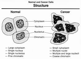
In this unit, we focused on the body's defense. Externally, our body has a first line of defense: the skin, which serves as a physical barrier against pathogens. Comprised of the epidermis, dermis, and hypodermis layers (outermost to innermost layers), the skin also uses chemical methods of preventing pathogens, such as secreting sweat, mucus, and its levels of keratin to resist bacterial enzymes. These forms of resistance are called non-specific. Other forms of non-specific resistance are other chemical methods inside the body like gastric juice, anti-microbial substances like natural killer cells, and anti-microbial processes like inflammation and fever. Other forms of resistance used by the body to defend against pathogens are called specific. These are cell-mediated or antibody-mediated responses that occur when the first layers of defense have been penetrated. Cell-mediated immunity uses T-cells, which can be either Helper T-cells, Cytotoxic T-cells, or Memory T-cells. B-cells make up the antibody-mediated immunity, developing into plasma cells and using neutralization, immobilization, agglutination, activation of complement, attraction of phagocytes, or phagocytosis enhancement to stop the threat. Generally, our bodies immune system kills "bad" cells like cancer cells in under 100 seconds (McCarthy). Sometimes, they don't neutralize the cancer cells, which then grow into tumors, masses of extra tissue. Cancer is the uncontrolled growth or body cells that crowd out normal cells. Some tumors are non-cancerous or benign, but cancerous, or malignant, tumors cause death if untreated. If uncontrolled, cancer cells will spread across the body in a process called metastasis. Cancer treatments include chemotherapy, removal surgery, and radiation.
This unit, we had the interesting opportunity to participate in a debate. The debate was informative and fun, albeit stressful. Our topic, the organ market, was unique because both sides were debatable and strong. I honestly wasn't leaning one way or the other on the topic, and I was very surprised how the audience was so staggered. My team worked very hard, and I was so proud of our efforts.
Works Cited
http://preventdisease.com/news/14/020314_Scientists-Find-That-The-Human-Body-Kills-Spontaneous-Cancers-Daily.shtml
 https://upload.wikimedia.org/wikipedia/commons/5/5d/Anatomy_The_Skin_-_NCI_Visuals_Online.jpg
https://en.wikipedia.org/wiki/Cancer_cell

Tuesday, November 8, 2016

Sheep Heart Dissection Questions

1. The pericardium serves to provide lubrication for the heart and protects against infection. It is the outermost layer of the heart.

2. The biggest difference between veins and arteries is that arteries take blood away from the heart, while veins bring blood to the heart. Veins have valves that prevent back-flow of blood. The veins also have thinner walls. Arteries have thick, muscular walls.

3. The auricles are like a balloon attached to the atria. They fill up with blood also, increasing the holding capacity of the atria.

4. The external structure gets narrower as we go down the heart. The atria are wider than the ventricles. This gives the appearance of the upside down triangle sort of shape.

6.

7. The chordae tendinae prevent the valve from reopening in response to the pressure lowering in the atrium during ventricular systole. The pressure in the atria becomes much lower, and because things naturally go from high to low concentration, the chordae tendinae and papillary muscles prevent this from happening.

8. The valve is a whitish color, different in texture from the endocardium lining of the chambers. This valve is bigger than the semilunar valves and the tricuspid valve.

9. Semilunar valves prevent back-flow from arteries back into the ventricles during ventricular diastole, and they also maintain the pressure in the arteries so they can effectively push the blood where it needs to go.

10. a) Blood can become backed up in other parts of the body if there is valve disease, causing swelling. If the blood can't get pumped back into the heart, it becomes stuck. b) Left side valve disease can cause tricuspid regurgitation.

11. The chordae tendinae of the bicuspid valve are super stringy, pulled taut with probe causes valve to be open. You can see through the aorta, or where the aortic arch would be.

12. The right side of the heart receives oxygen deprived blood and sends it to the lungs for reoxygenation. Conversely, the left side of the heart receives the oxygen rich blood from the lungs and sends it to the rest of the body through the left ventricle.


13.

Unit 3 Reflection

Review: This unit was about cardiovascular health, and how to maintain said health. The circulatory or cardiovascular system is comprised of three parts, the heart, the blood, and the blood vessels. The heart is the basis of operations: all blood in the body comes back to the heart to be reoxygenated and resent back to the body. The structure of the heart plays an important role in its function: four separated chambers operate through contraction and relaxation, called systole and diastole respectively, which pushed the blood out of arteries, particularly the pulmonary arteries and the aorta. The right side of the heart gets deoxygenated blood and sends it to the lungs, and the left side gets oxygenated blood and sends it to the body. The blood itself is comprised of different types of cells and fluid: red blood cells, white blood cells, plasma, and platelets. Blood vessels, like arteries, veins, and capillaries, transport the blood where it needs to go in the body.
Heart disease is caused by atherosclerosis, triggered by excess sugar, excess polyunsaturated fats, excess omega-6, and damaged blood vessels. Focusing on the latter, blood vessel damage occurs when LDL, the "bad cholesterol," sticks to the walls of the blood vessels, lodging itself in the walls. When this occurs, an inflammatory response is triggered, which results in a counterproductive process of creating "foamy" mast cells, creating a plaque in the walls. This plaque can break, which causes a blood clot. This blood clot can either block the blood in the same blood vessel, or break off and travel further in the body, where it clogs up there and causes a heart attack or a stroke. A stroke only occurs in the brain, and can be very fatal. Every minute during a stroke, 2 million blood cells die, making the treatment for strokes very time-sensitive. To promote your own cardiovascular health, maintain a healthy diet, exercise, do not smoke or drink alcohol in excess, and know your family history of hypertension.
Reflect: I am still struggling with differentiating between the different white blood cells. I struggled with the case study activity, which is concerning because that question format is a large portion of the test. Also, without my group members to collaborate with, I probably cannot identify structures on the sheep heart alone. If I had to look at another sheep heart that wasn't the one I was familiar with and hands-on with, I would be beyond confused.
Considering that it is currently past midnight and I'm still mid-study session, my sleep goal from last semester is not going so well. However, I was able to catch up on some of the midterm-stolen sleep over the weekend! These couple of weeks are extremely tough as many deadlines overlap. Most of the units in our various classes tend to end on a similar schedule, meaning that we have weeks of "chill" time and weeks of testing mayhem. This makes it hard as a student to allocate my time to all of my classes appropriately, and maintain my work schedule. In general, I'm a focused person, so I know that it's not lack of focus contributing to any poor marks, but rather just lack of time or biological ability to function. I do try as hard as I can.
Going back to the unit, a surprisingly fun activity was the dissection! I entered the activity with a negative mindset, intending to only take notes, but I was soon "two-glovin' it" and fully immersed, poking and prodding the heart with gusto. I'm happy with myself for pushing my learning to another level, despite my prior reservations.

Thursday, October 27, 2016

Mid-Unit Summary

This unit, we are learning about the circulatory system. The heart of the circulatory system is, metaphorically and literally, the heart itself.
Let's look at the structure of the heart. Comprised of four chambers, the heart is hollow to allow for blood to be pumped in and out. The chambers are the left ventricle, right ventricle, left atrium, and right atrium. The atria are the upper chambers, and the ventricles are below. The right atrium receives oxygen deprived blood. The left atrium receives oxygen rich blood. The right ventricle pushes blood out of the heart to the lungs. The left ventricle discharges blood to the body. There are valves in the heart that prevent back-flow of blood: the sublunar pulmonary and aortic valves, the tricuspid valve, and the bicuspid valve. The heart sounds we hear are the slamming of these valves shut.
The circulatory system branches from the heart to the entire body. The 3 components are the heart, the network of tubes, and the blood. Blood flows in veins to the hearts, or arteries away from the heart. Small arteries are called arterioles, and small veins are called venules.
Blood itself is comprised of 55% plasma and 45% cellular stuff. The cellular stuff is red blood cells, white blood cells, platelets, and others. The white blood cells are neutrophils, basophils, eosinophils, lymphocytes, and  monocytes. Platelets help blood clot. Red blood cells carry oxygen in the blood.
When there is an excess amount of LDL, the bad cholesterol, it sticks to artery walls, becoming a plaque, which can then rupture and cause a blood clot. Blood clots can lead to strokes and heart attacks, which are very deadly.

https://upload.wikimedia.org/wikipedia/commons/e/e0/Heart_diagram-en.svg
https://www.google.com/url?sa=i&rct=j&q=&esrc=s&source=images&cd=&ved=0ahUKEwiVstrt8PvPAhUQ9mMKHX-6A8QQjRwIBw&url=https%3A%2F%2Fen.wikipedia.org%2Fwiki%2FWhite_blood_cell&psig=AFQjCNG_kW_UUtA4aTszmYdRUT-yfK7fWA&ust=1477688519713371

Friday, October 21, 2016

Blood Pressure Lab

Systole is the pressure of the blood in the body when the heart contracts, while diastole is the remaining pressure when the heart is relaxed. To measure heart rate, you can use a blood pressure cuff and a stethoscope, but you can also use a timer and your fingers placed on the radial or carotid arteries. Blood pressure is measured with a blood pressure cuff and a stethoscope. When you measure a pulse, don't use your thumb because your thumb has its own independent pulse that you may feel. To use a blood pressure cuff, place the cuff, with no air, on the bicep area of the uncovered arm. Put the stethoscope end under the cuff, on top of your veins in the inside of your elbow. Close the valve on the cuff and squeeze the bulb until the meter of the sphygimomanometer says 150 mm/Hg. Gently release the air from the valve, listening for the first heartbeat. Take note of the number on the meter when you hear it. Continue releasing the air until you no longer hear the heartbeat, and take note of the number on the meter when you stop hearing the heartbeat. The first number recorded is the systolic number, and the second is the diastolic.